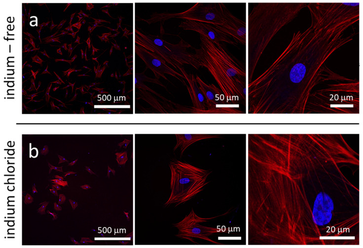
Figure 2.
Fluorescence confocal micrographs of adherent cells in petri dish prepared with (a) indium-free media and (b) media containing 3.2 mM of InCl3. Images show cell density decreases after they are exposed to indium chloride. Cells also become more compact and circular in shape. DNA molecules are visualized with DAPI in blue; actin filaments are stained with deep red Cytopainter F-Actin.

