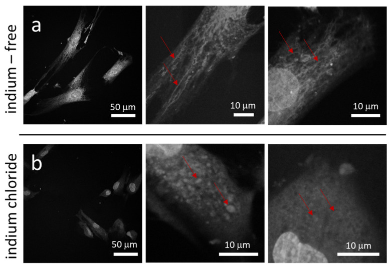
Figure 3.
Fluorescence confocal micrographs of mitochondria within adherent cells in culture dish prepared in (a) medium without InCl3 and (b) medium containing 3.2 mM of InCl3. Images show long tubular mitochondria form networks in cells incubated in indium-free media. When treated with indium chloride, mitochondria are punctate in shape. The red arrows highlight stained mitochondria.

