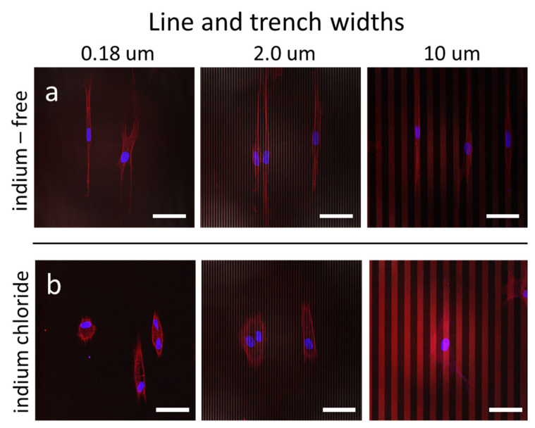
Figure 4.
Fluorescence confocal micrographs of adherent cells on parallel line/trench structures incubated in (a) medium without InCl3 and (b) medium containing 3.2 mM of InCl3. Images show cells elongated and aligned to the line axes when cultured in baseline media without indium chloride. After exposure to the indium chloride, cells are more compacted and less likely to align to the pattern axes. Scale bars correspond to 50 µm. Cell nuclei appear blue (4′,6-diamidino-2-phenylindole; DAPI), whereas F-actin microfilaments appear red (fluorescent phalloidin conjugate).

