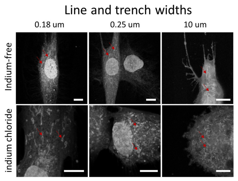
Figure 5.
Fluorescence confocal micrographs of adherent cells stained with MitotrackerTM on parallel line/trench structures with widths of 0.18 µm, 0.25 µm and 10 µm. Cells were cultivated in two different medium compositions—without and with indium chloride. Scale bars correspond to 10 µm. The red arrows highlight some of the mitochondria.

