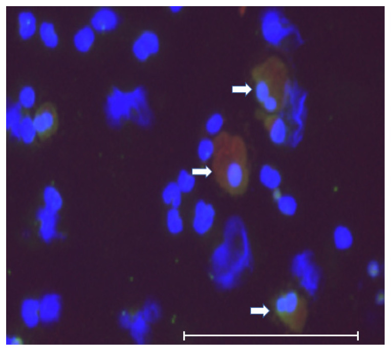
Figure 4.
Morphology of double positive cells. These cells were large, polymorphic, and polynuclear, which suggested that they were tumor–macrophage fusion cells (TMFs), macrophage–tumor cell fusion cells (MTFs), or cancer-associated macrophage-like (CAMLs) cells. The scale bar shows 150 μm. White arrows indicate CK/CD45 double positive cells.

