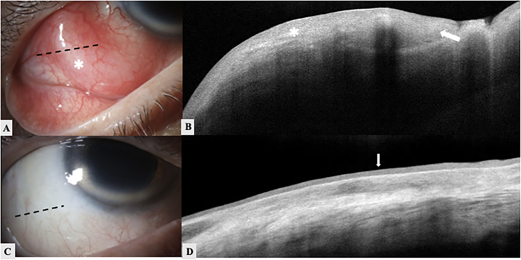
Figure 2. An 82-year-old white male with an invasive squamous cell carcinoma (SCC) of conjunctiva of the right eye.

A. Slit lamp image of the right eye shows an immobile elevated nodular conjunctival lesion (white asterisk) with feeder vessels from 7 to 9 o’clock. The black dotted line demonstrates the orientation of the HR-OCT raster. B. HR-OCT showing marked thickened hyperreflective epithelium (asterisk) with significant posterior shadowing effect. Invagination of abnormal epithelium is noted (white arrow). C. Slit lamp image of the same patient after surgical treatment and brachytherapy. The black dotted line demonstrates the orientation of the HR-OCT raster. Excisional biopsy confirmed invasive SCC. D. HR-OCT of the same eye after treatment showing normal thickness epithelium with slight hyperreflectivity (white arrow). The mass is resolved.
