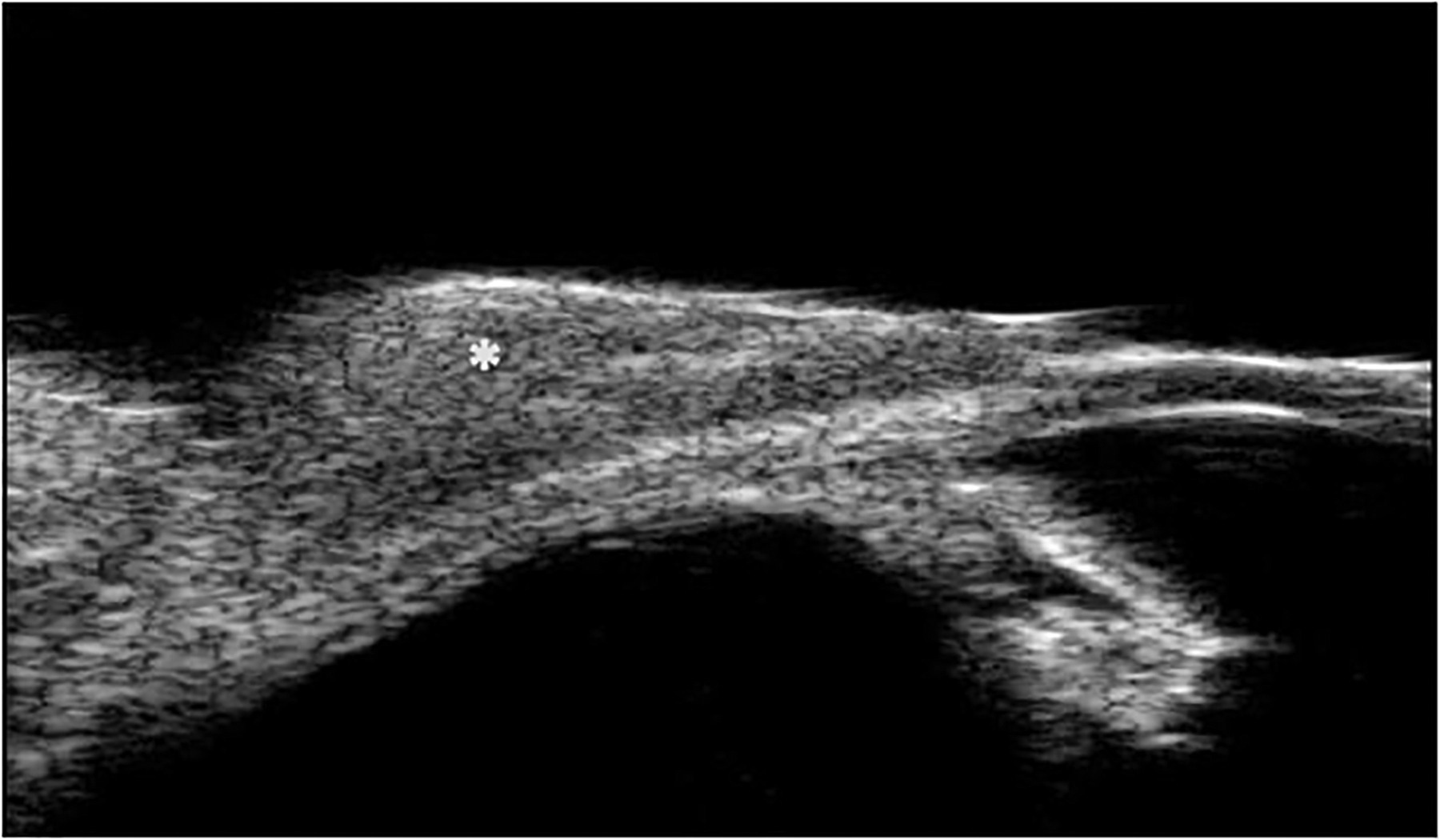
Figure 4:

High-resolution ultrasound biomicroscopy (UBM) image shows a dome-shaped, non-vascularized, slightly hypoechoic lesion over the sclera (asterisk). No definite scleral involvement is evident on the UBM. However, the clinical examination and final pathologic report confirmed the scleral invasion of this SCC lesion.
