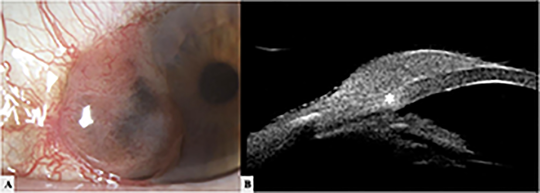
Figure 7: A 90-year-old white female with conjunctival melanoma on the right eye.

A.Slit lamp image shows a highly elevated mixed melanotic and amelanotic lesion on the bulbar conjunctiva and abutting the cornea from 7–10 o’clock with feeder vessels. B. High resolution UBM image shows a dome-shaped, regular structure with a mild hypoechoic core. Possible invasion into the corneal limbus at the 9 o’clock position (asterisk) is noted.
