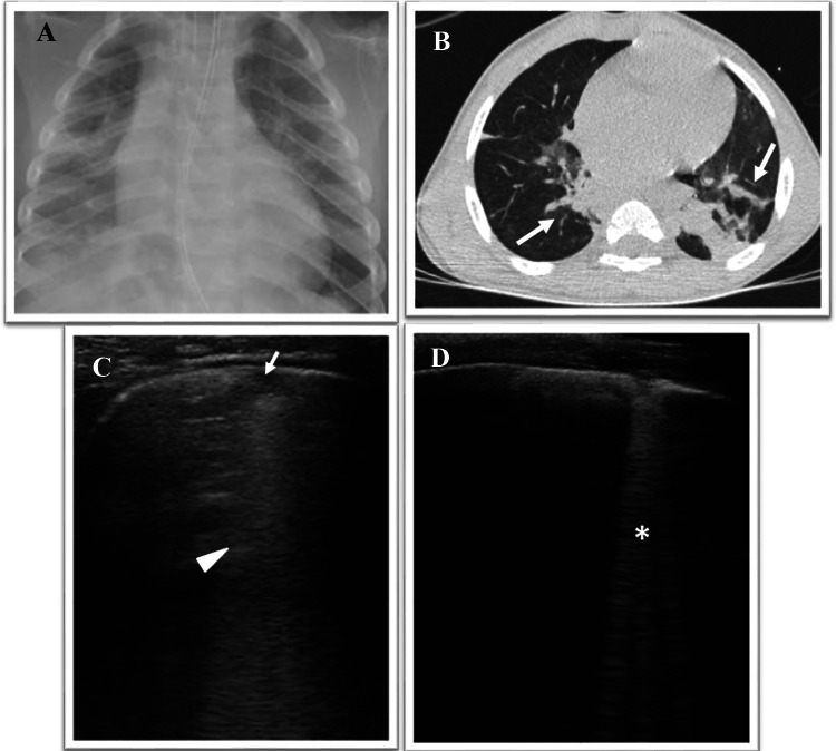
Fig. 4.
a Chest digital radiography shows areas of HyperLucency in the middle and inferior right lung fields and in the lower left lung fields consisting with consolidation. b Axial CT image shows bilateral subsegmental ground glass opacities with partial consolidation in posterior segments of the lungs (gravity dependent) (arrow), consisting with aspiration pneumonia. c, d Grayscale lung ultrasound examination during the follow-up, shows: disappearance of subpleural consolidations that give way to ultrasound interstitial syndrome characterized by the bilaterally presence of—at first c—areas of subpleural micro consolidations (arrow) associated with “white lung” and coalescent B-lines (arrowhead) and—subsequently, in the resolution phase d—areas of single and non-coalescent B-lines (asterisk)

