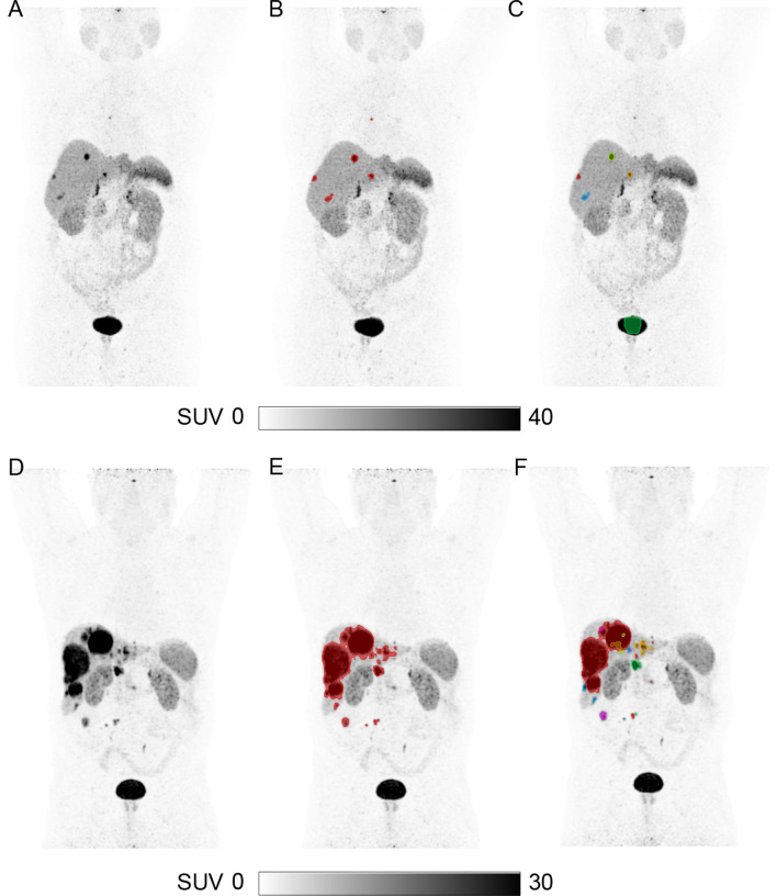
Fig. 3.
Representative examples of the segmentations from the AI model for two patients. Maximum intensity projection [64Cu]Cu-DOTATATE PET without tumor segmentation (A, D). Ground truth segmentation of tumor (B, E). AI predicted segmentations—no manual adjustments performed (C, F). In the AI output, all separate segmentations are given a unique color, e.g., red, blue, green, making manual adjustment with deletion of a segmentation easy and fast (e.g., part of the bladder was erroneously segmented in C)

