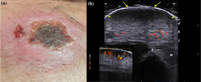
Fig. 1.
a) Clinical and sonographic presentations of an 18-year-old male presenting an LCL plaque with a crusted center and raised margins, located on the right elbow. b) Greyscale ultrasound scan at 18 MHz detected an ill-defined dermo-hypodermal structure with mixed echogenicity (between yellow markers) extending up to the fascial plane. Note the loss of epidermal layer (yellow arrows), the increased echogenicity of fatty lobules (red markers), and the dilation of interlobular spaces (red arrows). PDUS Doppler in the bottom left box shows a prominent vascular signal in the center of the lesion, consistent with the inflammatory pattern

