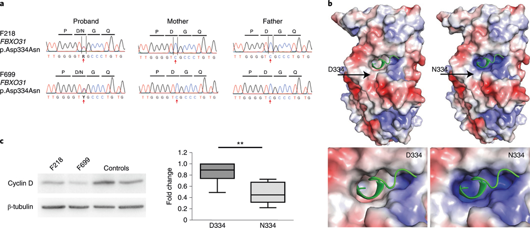
Fig. 2 |. Functional validation of the CP-associated FBXO31 variant p.Asp334Asn shows alterations in cyclin D regulation.
a, Sanger traces of the mother, father and proband from families F218 and F699 verify de novo inheritance and the position of the variant (red arrow). b, Poisson–Boltzmann electrostatic maps of wild-type FbXO31 (left) and the p.Asp334Asn variant (right). D334 is positioned around the cyclin D1 (green)-binding pocket on FbXO31. the mutation alters the surface electrostatic charge around the cyclin D1-binding site with a predicted effect on cyclin D1 binding to FbXO31. the site of D334/N334 has been labeled (arrow). the bottom panels are magnified views showing the alterations to the surface charge in the cyclin D1-binding site. c, A representative western blot cropped to show the decreased cyclin D expression in patient-derived fibroblasts with the FBXO31 p.Asp334Asn variant. Quantification of cyclin D is normalized to in-lane β-tubulin and the within-experiment control GMO8398. both patients had reduced cyclin D compared to pooled controls. the data are averaged for three independent cell culture experiments (n = 7 controls, n = 6 patient measurements). the box indicates the 75th and 25th percentiles with a center line indicating the median; the whiskers indicate the 10th and 90th percentiles. **P = 0.004 calculated using a two-tailed unpaired t-test. Full-length blots are provided as source data.

