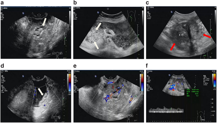
FIG. 1.
Conventional ultrasonic characteristics of OSSPBT. (a) A large area of solid echo was seen around the ovary, and a normal ovary with small follicles (white arrow) was seen in the mass. (b) The ovary is surrounded by a large number of solid mixed echoes, and a large number of speckled strong echoes are seen, showing “blizzard” sign (white arrow). (c) MOSC was bilateral ovarian irregular solid or mixed echo mass (red arrow), no normal ovarian tissue echo. (d) There were tumor planting nodules (white arrow) and punctate blood flow signals in the retroperitoneum of the pelvic cavity. (e, f) CDFI showed mild or moderate blood flow signals in the tumor, PSV: 10.9 cm/s, RI: 0.41, and color score: 3. CDFI, color doppler flow imaging; LM, left ovarian lesion; Mass, lesions; MOSC, malignant ovarian serous cystadenocarcinoma; OV, ovary; PSV, peak systolic velocity; RI, resistance index; RM, right ovarian lesion; UT, uterus.

