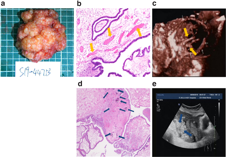
FIG. 3.
Pathological image of OSSPBT and its corresponding ultrasonic image. (a) The gross appearance of the tumor was a large number of white and transparent bead-like nodules fused into a cauliflower pattern. (b) HE staining showed the papilla or micropapillary structure of the cells covered with single layer or multilayer cubic to columnar cells, and there were more dilated and congested microvessels (yellow arrows) in the axis of connective tissue ( × 200). (c) The irregular branching blood vessels (yellow arrows) were visible in its corresponding 3D ultrasound image. (d) HE staining showed that the fiber connective tissue axis saw a large amount of gravel (blue arrows) ( × 200). (e) A large number of strong echo speckled echo spot (blue arrows) in the tumor were visible in its corresponding conventional ultrasound image. HE, Hematoxylin–Eosin.

