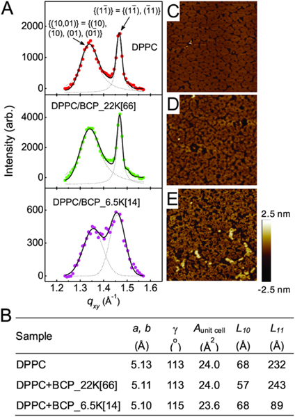
Figure 4.
Effects of BCP_22K[66] and BCP_6.5K[14] on packing of lipids. (A) Bragg peaks from GIXD on a pure DPPC monolayer (top panel), and a DPPC monolayer in the presence of BCP_22K[66] (middle panel) or BCP_6.5K[14] (bottom panel) in a water subphase at π = 30 mN/m and T = 23 °C. The two Bragg peaks observed from the DPPC monolayers indicate distorted-hexagonal packing of the lipid tails in a 2-D unit cell and the corresponding Miller indices are indicated for each peak. (B) Parameters obtained from the GIXD profiles. a, b, and γ are the axes of a unit cell and the angle across from the a and b axis, respectively; Aunit cell is the area of a unit cell; L10 and L11 are coherence length in the direction of {(10, 01)} and {11}, respectively. The GIXD measurements were taken at ADPPC = 47.3 Å2, ADPPC+BCP_22K[66] = 47.3 Å2 and ADPPC+BCP_6.5K[14] = 61 Å2. (C−E) AFM height images (2 μm × 2 μm) of the DPPC monolayer (C), DPPC monolayer in the presence of BCP_22K[66] (D), or BCP_6.5K[14] (E) on a water subphase deposited onto a mica substrate at π = 30 mN/m and T = 25 °C. The two different domains observed in the DPPC monolayers are the liquid condensed (LC) phase (bright pinwheel-like shape) that gives rise to Bragg diffraction in GIXD, and the liquid-expanded (LE) phase (darker noncontinuous area) within which the hydrocarbon chains are highly disordered.

