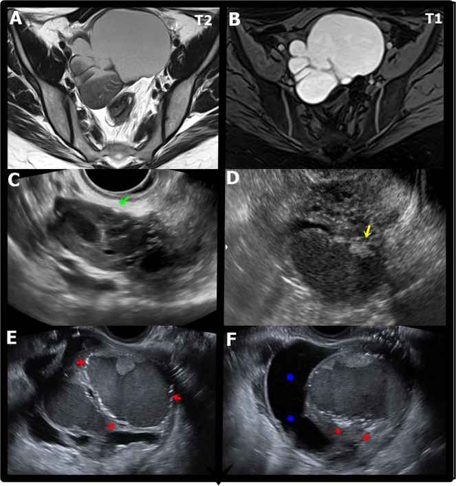
Figure 1.

MRI and ultrasound appearances of ovarian endometriomas. A. Axial T2W MR image shows a multiloculated endometrioma with variable appearances of “T2 shading”. B. Axial T1W MR image with fat saturation shows a multiloculated endometrioma with marked and uniform hyperintensity with a “light bulb bright” appearance. C. Transvaginal ultrasound (TVUS) shows a small endometrioma with small echogenic focus in the ovarian parenchyma (green arrow).D. TVUS) shows an ovarian endometrioma with heterogeneous content due to small papillary projection (yellow arrow). E. TVUS shows two ovarian endometriomas, the largest with irregular contours and hyperechogenic focus in the wall (red arrows).F. TVUS showing an ovarian endometrioma with irregular contours and hyperechogenic focus in the wall (red arrows) associated with paraovarian inclusion cyst seen as an indirect sign of adherence process (blue dots).
