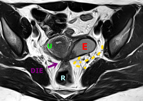
Figure 3.

Axial T2W MR image shows a deep endometriosis (DE) plaque in the posterior uterus with adhesions that extend from the torus uterus, invading the wall of the rectum and promoting retraction and medialisation of the left ovary that contains endometrioma (E). Bowel-invasive endometriosis of the rectum is also present with a “mushroom cap” lesion. U: Uterus, E: Endometrioma, R: Rectum, DIE: DE plaque.
