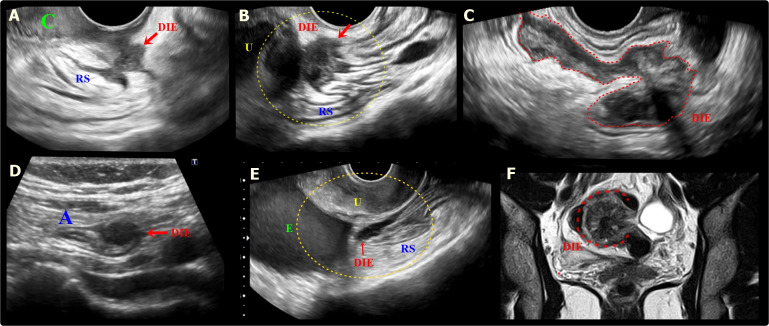
Figure 4.
A. Sagittal transvaginal ultrasound (TVUS) image shows a hypoechoic endometriotic nodular lesion, with irregular margins, infiltrating the low retrocervical space, with extension to the posterior vaginal fornix and the serosa of the anterior rectal wall (RS) (red arrow). DIE: deep endometriosis. B. Sagittal oblique TVUS image shows a hypoechoic lesion (red arrow) with ill-defined margins, covering and infiltrating the posterior uterine wall and the anterior rectosigmoid wall (RS), causing retraction and angulation of this segment. U: Uterus. C. Sagittal oblique TVUS image shows a large hypoechoic endometriotic lesion in plaque (red dashed) infiltrating the anterior wall of the rectosigmoid colon. D. TVUS image demonstrates a hypoechoic nodule (red arrow) infiltrating the appendiceal tip (A). E. Sagittal oblique image of TVUS shows a hypoechoic endometriotic lesion infiltrating the retro- and paracervical space, with extension to the anterior wall of the rectosigmoid colon (RS) (red arrow). Also note, the presence of an ovarian endometrioma (E). U: Uterus. F. Coronal T2W MR image shows deep endometriosis lesion with archiform morphology in the rectosigmoid (red dashed).

