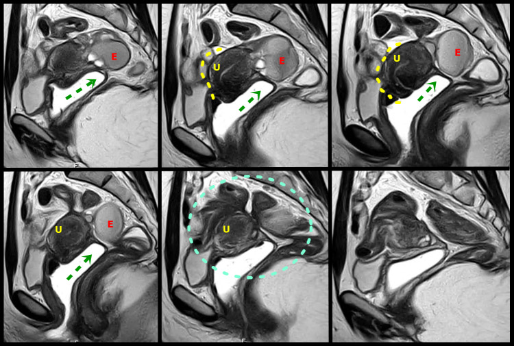
Figure 5.
Sagital T2W MR images with distension of the vagina by aqueous gel show a plaque in the rectouterine and retrocervical space, invading and elevating of the posterior vaginal fornix (green arrow), causing obliteration of the posterior cul-de-sac (green circle). The lesion infiltrated the posterior uterine serosa and myometrium and is causing uterine retractile retroflexion (yellow interrupted line)A left adnexal endometrioma (E) demonstrates “T2 shading” and associated fibrotic change. U uterus.

