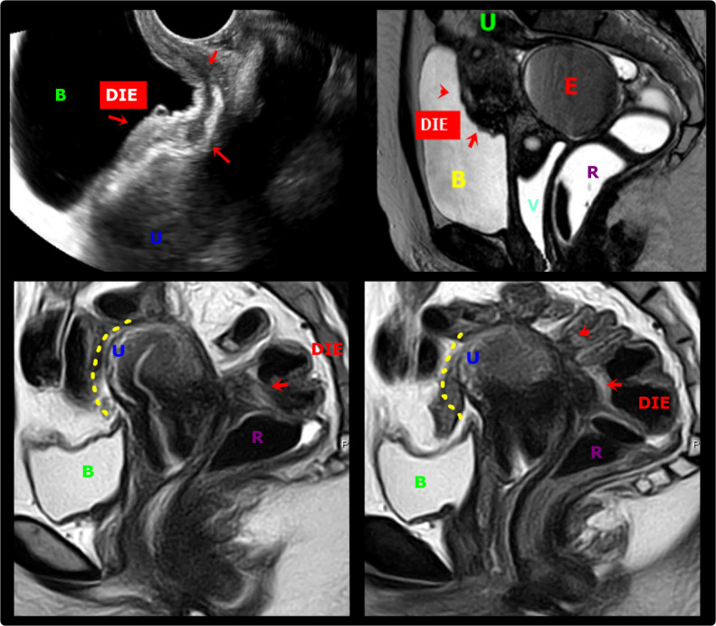
Figure 6.

A & B. Transvaginal ultrasound and axial T2-weighted MR images show deep endometriosis (DIE, red arrows) obliteration of the peritoneum of the vesico-uterine space with involvement of the posterosuperior bladder wall. The lesion infiltrates the peritoneum of the vesicouterine space, causing the obliteration of the fatty planes between adjacent structures. B: Bladder, U: Uterus, R: Rectum, E: Endometrioma, V: Vagina; C & D. Sagittal T2W MR images show a deep endometriosis (DIE) plaque in the retrocervical space with bowel involvement, causing obliteration of the posterior cul-de-sac with loss of fat planes between adjacent structures and forced retroflexion of the uterine fundus (yellow interrupted line). B: Bladder, U: Uterus, R: Rectum, E: Endometrioma
