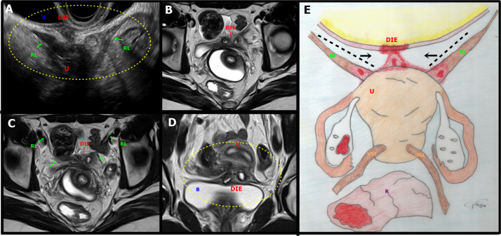
Figure 7.
A. Sagittal oblique transvaginal ultrasound image shows a hypoechoic endometriotic lesion with irregular and ill-defined margins that infiltrated and obliterated the peritoneum of the vesicouterine space, extending to the insertion of the round ligaments (RL), more evident on the right. B: bladder, DIE: deep endometriosis, U: uterus, RL: round ligament. B-D. Axial (B, C) and coronal (D) T2-weighted MR images show irregular thickening and medialisation of the round ligaments (low signal intensity) at their insertion sites, near the uterus. Focal thickening of the detrusor muscle of the bladder and the anterior uterine serosa is also seen. Abbreviations as in A. E. Illustrative figure showing a focus of deep endometriosis (DIE) in the anterior compartment of the pelvis. R: Rectum, U: Uterus, RL: Round ligament, USL: Uterosacral ligaments.

