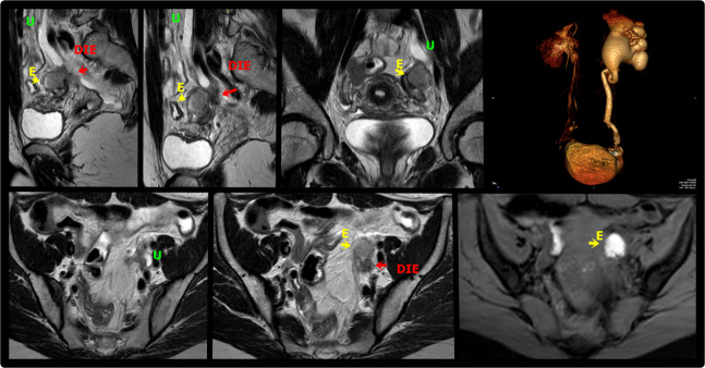
Figure 8.
Sagittal (A, B), coronal (C) and axial (E,F) T2W and axial T1W with fat saturation (G) MR images demonstrate deep endometriosis thickening of the left paracervical area(arrow) involving the parametrium and extending to the ovarian fossa and adjacent ureter, resulting in upstream hydroureter and hydronephrosis. There is also an endometrioma in the left ovary (E). MR urogram volumetric reconstruction shows asymmetry of the ureters with accentuated hydronephrosis to the left (D). DIE: deep endometriosis, U: Ureter, E: Endometrioma.

