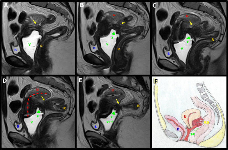
Figure 9.
A-E. Sagittal T2W MR images with distension of the vagina (V) by aqueous gel show a stellate low-signal-intensity endometriotic lesion containing small cystic areas in the retrocervical space, with indirect signals of adherence; infiltration of the bilateral uterosacral ligaments, elevation of the posterior vaginal fornix (green arrow),causing obliteration of the posterior cul-de-sac and uterine retractile retroflexion (red interrupted line). Bowel-invasive endometriosis of the rectum is also present (yellow arrows). F. Schematic representation of the imaging signs shown in A-E sequences: Vagina, C: Cervix, U: Uterus, B: Bladder, RS: Rectosigmoid, PF: Posterior Fornix.

