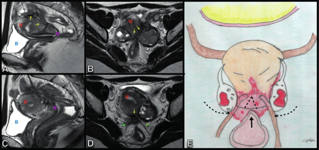
Figure 10.

Sagittal (A,C) and axial (B, D) T2W MR images show a deep endometriosis lesion in the retrocervical space, in the topography of the uterine torus (yellow arrow) and insertion of the uterosacral ligaments (green arrow) with extension to the peritoneum of the ovarian fossae with posteriorisation and medialisation of the ovaries (“Kissing ovaries”). There is also extension of the lesion to the rectosigmoid colon with cranial angulation and retraction in the loop segment involved. DIE: Deep Endometriosis, U: Uterus, B: Bladder.
