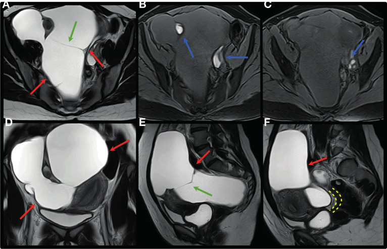
Figure 13.
Axial, coronal and sagittal T2W (A, D, E, F) and axial T1W MR images with fat saturation (B,C) show large cyst of peritoneal inclusion occupying practically the entire pelvic cavity (red arrows), with some fine septations (green arrows), bilateral ovarian endometriomas (blue arrows) and deep endometriosis infiltrating the anterior rectosigmoid wall (yellow interrupted line).

