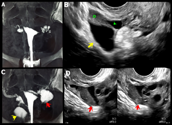
Figure 14.

A, C. Hysterosalpingography showing indirect signs of adhesion process in both adnexal regions, characterised by absence of free distribution of the contrast (yellow and red arrows). B. Axial oblique transvaginal ultrasound (TVUS) image demonstrating right para-ovarian peritoneal inclusion cyst with fine septum (green arrow).D. Axial oblique TVUS image shows a hypoechoic endometriotic lesion infiltrating the left ovarian fossa (red arrows) associated and loculated fluid in this location.
