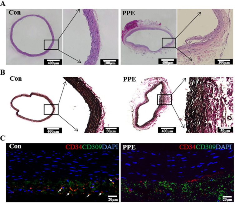
Fig. 3.
Histological sections of abdominal aorta. (A) Hematoxylin and eosin staining of the aorta. (B) Verhoeff-Van Gieson (VVG) staining of elastic fibers in aneurysm sections. (C) Immunofluorescence of abdominal aorta sections labeled with antibodies to detect CD34 (red) and CD309 (green). The white arrow indicates CD34+/CD309+ cells colocalizing as orange.

