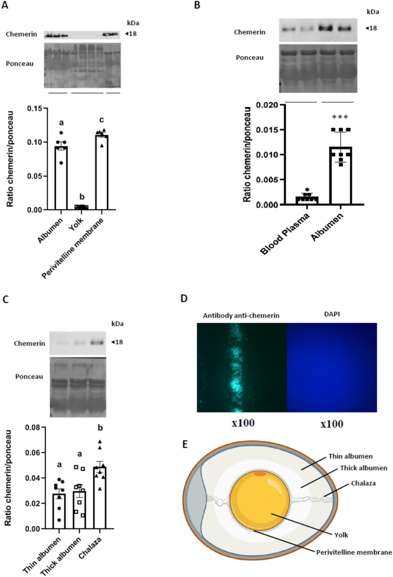
Figure 1.
Chemerin accumulation within the egg. Ten hens were randomly selected to collect blood plasma, and six eggs were randomly selected to sample thick and thin albumen, perivitelline membrane, yolk, and chalazas. (A) Protein abundance of chemerin detected by western blotting within the albumen (mix of thin and thick), yolk, and the perivitelline membrane in the eggs (n = 6). (B) Protein abundance of chemerin detected by western blotting within the blood plasma and the albumen (mix of thin and thick) of hens (n = 8 animals and eggs). (C) Protein abundance of chemerin detected by western blotting within the thin and thick albumen, and the chalaza of eggs (n = 10). (D) Immunofluorescence for chemerin qualitative detection within the perivitelline membrane (magnification ×100). (E) Localisation of different components (thin and thick albumen, yolk, chalaza, and perivitelline membrane. The values are expressed as mean ± standard errors of means. Different letters indicate significant differences at p < 0.05 and ***indicate significant differences at p < 0.001.

