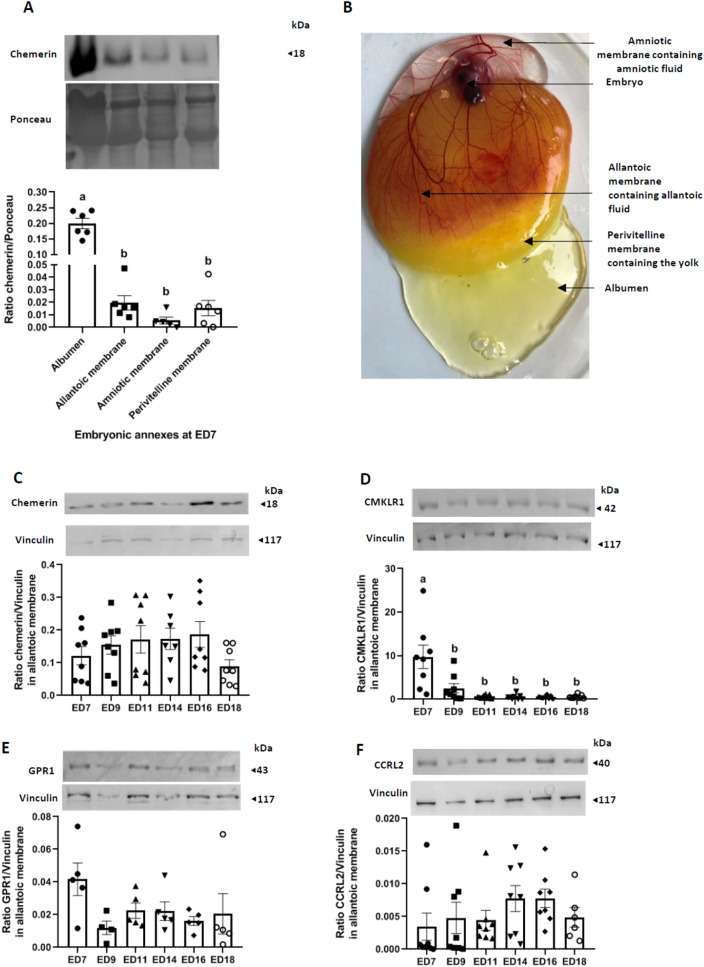
Figure 4.
Chemerin system expression in embryo annexes and fluids. (A) Protein abundance of chemerin detected by western blotting within the albumen (thin and thick), allantoic membrane, amniotic membrane, and the perivitelline membrane from fertilised eggs incubated for 7 days (n = 6). The ratio chemerin/ponceau is represented. (B) Localisation of the different embryonic structures at ED7. (C–E) Protein abundance of chemerin (C), Chemokine-like receptor 1 (CMKLR1) (D), G Protein-coupled Receptor 1 (GPR1) (E) and Chemokine (C–C motif) receptor-like 2 (CCRL2) (F) detected by western blotting within the allantoic membrane of incubated eggs at different embryonic days (ED): 7, 9, 11, 14, 16, and 18 (n = 8 per stage). Values are expressed as mean ± standard errors of means. Letters indicate significant differences between various conditions (p < 0.05).

