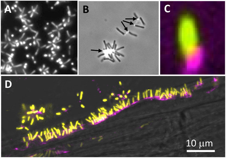
Figure 1. UPP-mediated polar attachment in A. tumefaciens.
A. tumefaciens UPP on abiotic surfaces and plant tissues. Fluorescence microscopy using wheat germ agglutinin lectin labeled with FTIC (fl-WGA) and Alexa Fluor 594 (af-WGA). (A-C) Attachment assays on 22 x 22 mm glass coverslips. A. tumefaciens C58 cells incubated for the indicated time in ATGN minimal medium with coverslips prior to lectin labeling with 10 μg/ml of either fl-WGA or af-WGA for 20 min. (A) fl-WGA labeling of a 12 h biofilm. Merged image from bright field and fluorescence microscopy. (B) af-WGA-labeling of a short-term attachment assay after 2 h incubation. Merged image from phase contrast and fluorescence. (C) af-WGA labeling of a single A. tumefaciens cell attached to glass expressing plasmid-borne Ptac::gfpmut3 (pJZ383). Scanning Disk Laser Confocal fluorescence microscopy; Merged green and red channels, pseudo-colored yellow and magenta, respectively. (D) Binding of A. tumefaciens (pJZ383) to Arabidopsis thaliana WS 1 cm root cutting incubated for 3 h in ATGN minimal medium. Merged green and red channels pseudo-colored yellow and magenta, respectively plus bright field. Microscopes: Nikon E800 fluorescence microscope and Photometrics Cascade cooled CCD camera; Spinning Disk Nikon TE2000U microscope Yokagawa CSU10 confocal scanner unit.

