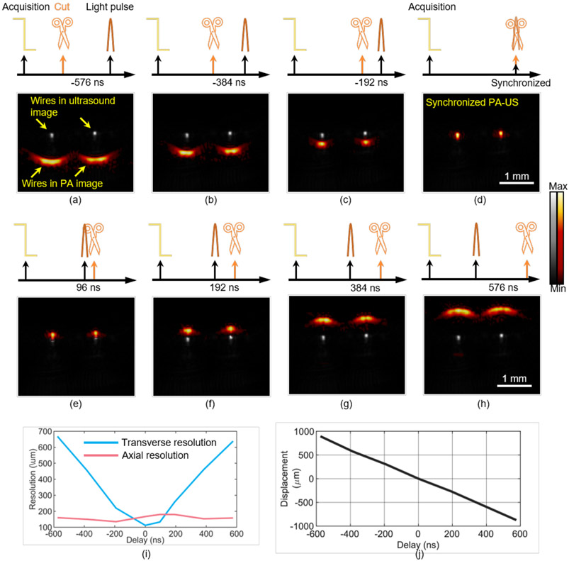
Fig. 3. Validating Scissors in ex vivo imaging.
Two nichrome wires were used as the sample. RF data are cut at different time with respect to laser pulses and PA-US images are acquired in real time (a–g): −576 ns (a), −384 ns (b), −192 ns (c), 0 ns (d), 96 ns (e), 192 ns (f), 384 ns (g), and 576 ns (h). Panel (i) shows the lateral resolution (blue line) and axial resolution (red line) when Scissors cuts RF data at different delays relative to the laser pulse. (j) The displacement of the left wire from its true depth in the PA image at different delays.

