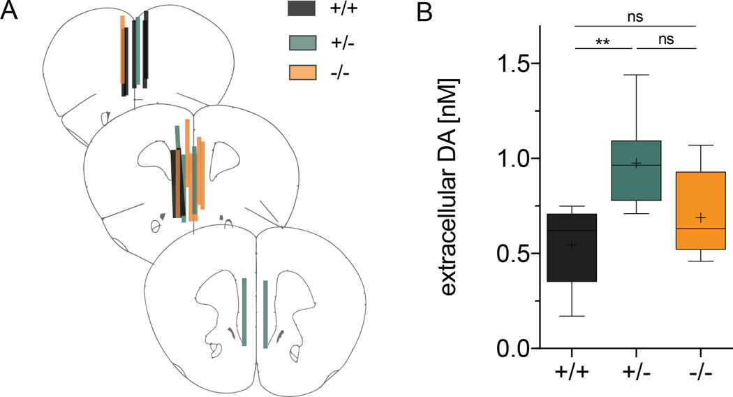
Figure 5. Tonic extracellular DA levels are increased in Cyt-1 heterozygote mice.
Extracellular DA levels were analyzed by unilateral no-net-flux microdialysis in the mPFC of freely moving control (black), heterozygote (green) and Cyt-1 KO (orange) mice. (A) Verification of microdialysis probe placement in the mPFC at bregma levels +2.22 mm, +1.98 mm and +1.78 mm in 50 μm-thick sections using Nissl staining. (B) Measurements for tonic extracellular DA levels in the mPFC of Cyt-1 mutants and controls (+/+ 0.546 ± 0.080 nM, +/− 0.977 ± 0.103 nM, +/+ 0.689 ± 0.085 nM, n=6–7/genotype, one-way ANOVA, F(2,17)=5.863, *p=0.0116; +/+ vs. −/− p=0.4876, +/+ vs. +/− **p=0.0094, +/− vs. −/− p=0.089).

