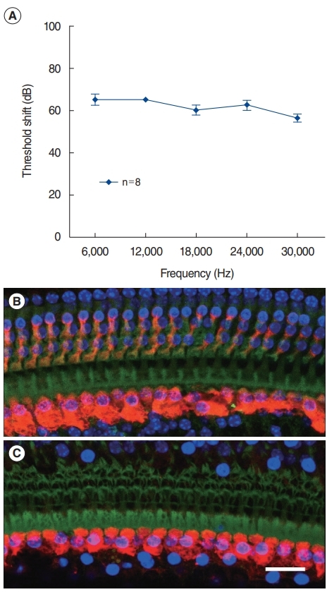
Fig. 1.

Permanent threshold shift. Mice were exposed to 120-dB white noise for 1 hour. The findings 2 weeks after noise exposure are shown. (A) Auditory brainstem response threshold shift. (B) The basal turn of the unexposed cochlea shows preserved inner and outer hair cells. (C) The noise-exposed cochlea shows degenerated outer hair cells and intact inner hair cells. The loss of nuclei (DAPI) and cell bodies (Myo7A) indicates degenerated outer hair cells. Only a cuticular plate is observed. Green, phalloidin; Blue, DAPI; Red, Myo7A. Scale bar=20 μm.
