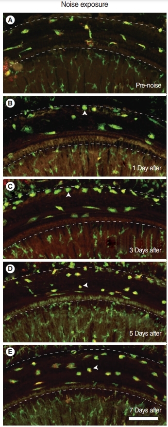
Fig. 4.

Migration of macrophages after acoustic injury. Whole mounted images of the basilar membrane using a confocal microscope at each time point are presented. Several CX3CR1+ resident macrophages are seen beneath the basilar membrane in (A) unexposed cochlea and (B) 1 day after noise exposure. (C) Three days postnoise exposure, an increase in CX3CR1+ cells with amoeboid morphology was seen in the junction of the basilar membrane and lateral wall (crista basilaris). Macrophages spread to the basilar membrane (D) 5, and (E) 7 days after noise exposure. All images show the lateral wall at the top and the osseous spiral lamina at the bottom. Arrowheads indicate representative amoeboid macrophages. Dotted lines indicate the borders of the basilar membrane. Green, CX3CR1; Red, F4/80; Scale bar=100 μm.
