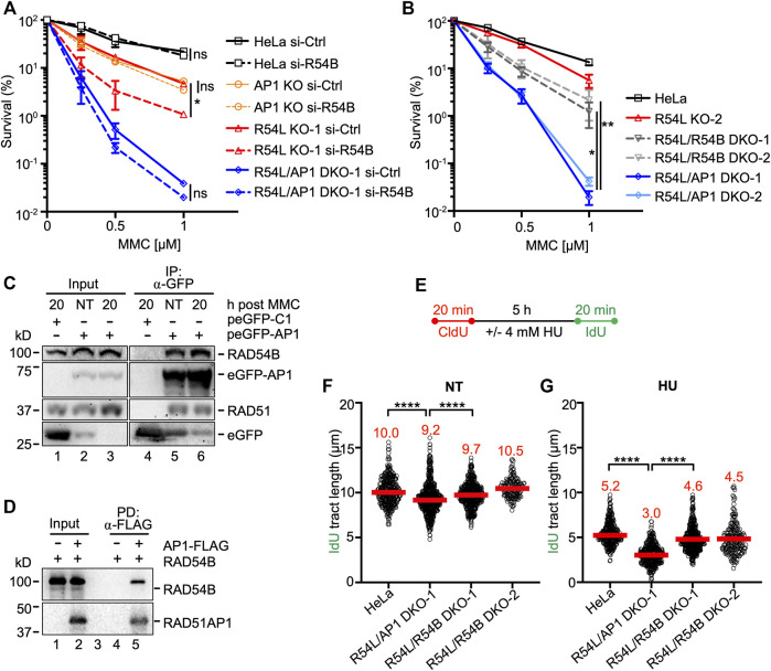
FIGURE 4.
Concomitant loss of RAD54L and RAD54B exacerbates MMC cytotoxicity and replication stress less severeley than concomitant loss of RAD54L and RAD51AP1. (A) Results from clonogenic cell survival assays after MMC of cells transfected with negative control (si-Ctrl) or RAD54B siRNA (si-R54B): HeLa, AP1 KO, R54L KO-1, R54L/AP1 DKO-1 cells. Data points are the means from two independent experiments ±SD. *, p < 0.05; ns, non-significant; two-way ANOVA followed by Tukey’s multiple comparisons test. (B) Results from clonogenic cell survival assays after MMC treatment of HeLa, R54L KO-2, R54L/54B DKO-1 and KO-2, and R54L/AP1 DKO-1 and DKO-2 cells. Data points are the means from two independent experiments ±SD. *, p < 0.05; **, p < 0.01; two-way ANOVA followed by Tukey’s multiple comparisons test. (C) Western blots to show that endogenous RAD54B co-precipitates in anti-eGFP protein complexes of R54L/AP1 DKO cells ectopically expressing eGFP-RAD51AP1 (here: peGFP-AP1) in the absence of MMC (NT; lane 5) and 20 h after a 2-h incubation in 0.5 μM MMC (lane 6). RAD51: positive control for interaction, as previously shown in different cell types (Kovalenko et al., 1997; Wiese et al., 2007). Lane 4: Neither RAD54B nor RAD51 co-precipitate in anti-eGFP protein complexes generated from R54L/AP1 DKO cells transfected with control plasmid (peGFP-C1). (D) Western blots to show direct interaction between purified FLAG-tagged RAD51AP1 protein (here: AP1-FLAG) and purified RAD54B precipitated by anti-FLAG M2 affinity resin (lane 5). (E) Schematic of the protocol for the DNA fiber assay. (F) Median IdU tract length under unperturbed conditions (NT) in HeLa, R54L/AP1 KO-1, and R54L/R54B DKO-1 and DKO-2 cells. Data points are from 150 to 200 fibers of three experiments for HeLa, R54L/AP1 DKO-1 and R54L/R54B DKO-1 cells and from one experiment for R54L/R54B DKO-2 cells, with medians in red. (G) Median IdU tract length after HU in HeLa, R54L/AP1 KO-1, and R54L/R54B DKO-1 and DKO-2 cells. Data points are from 150 to 200 fibers of three experiments for HeLa, R54L/AP1 DKO-1 and R54L/R54B DKO-1 cells and from one experiment for R54L/R54B DKO-2 cells, with medians in red. ****, p < 0.0001; Kruskal-Wallis test followed by Dunn’s multiple comparisons test.

