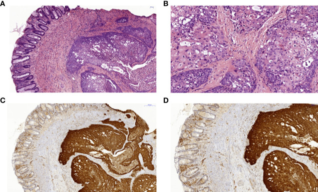
Figure 3.
Histopathology and immunohistochemistry of a squamous cell carcinoma of the anal canal in patient II.2. (A) Colorectal biopsy in H&E staining: left side colorectal mucosa, right side squamous cell carcinoma. (B) Anal squamous cell carcinoma in H&E staining with higher magnification with cellular atypia and dyskeratoses. (C) Anal squamous cell carcinoma showing positive p16 immunohistochemical staining of the tumor cells as a surrogate marker for HPV-infection in 7x magnification and (D) in 10.6x magnification. HPV, human papilloma virus.

