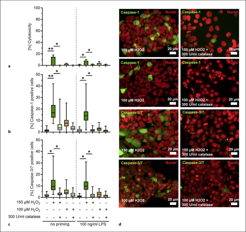
Fig. 4.
H2O2 activates caspase-1 and caspase-3/7 in bronchial epithelial cells. Unprimed or primed 16HBE cells were stimulated with 150 and 100 μM H2O2 in the presence or absence of catalase for 4 h and cytotoxicity (a) as well as caspase-1 and caspase-3/7 (b–d) activation was assessed. The stimulated cells were stained using fluorescent inhibitor probe FAM-YVAD-FMK (b, d) and FAM-DEVD-FMK (c, d) to microscopically visualize active caspase-1 and caspase-3/7, respectively. Nuclear-ID stain was used to visualize cell nuclei. Caspase-1- and caspase-3/7-positive cells were counted and are presented as a percentage of positive cells in relation to the total number of cells (b, c). Bars (a) denote mean values ± SD. The data in (b, c) are displayed as box plots. The level of significance was determined using Kruskal Wallis test with Dunn's post-test (n = 4; *p < 0.05; **p < 0.01; ***p < 0.001). SD, standard deviation.

