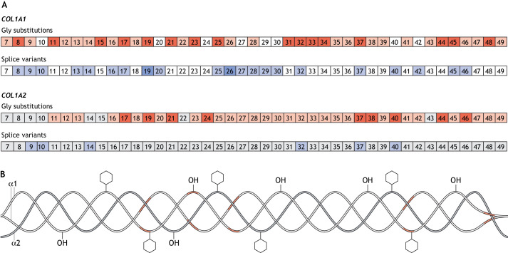
Fig. 1.
Lethal regions in collagen type I. (A) Exons containing glycine substitutions (red) and splice variants (blue) in COL1A1 (top) and COL1A2 (bottom) that are associated with phenotypic variability. Exons of which variants have been identified in >20 affected individuals are indicated by a darker shade of red and blue. Exons without reported sequence variants are in white for COL1A1 and in grey for COL1A2. (B) Structure of collagen type I protein showing the regions associated with lethal mutations in α1(I) in red.

