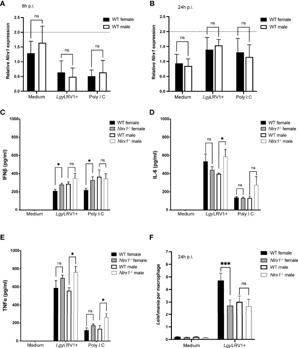
Figure 4.
Sex bias in inflammation and infection in absence of NLRX1. BMDMs from female and male WT and Nlrx1-/- mice were isolated simultaneously and infected with LgyLRV1+ parasites or stimulated with TLR3 agonist poly I:C (2 μg/ml). After 8 hours (A) and 24 hours p.i. (B), Nlrx1 mRNA levels were quantified by qRT-PCR (n=3 independent experiments). (C–E) After 24 hours, proinflammatory cytokines IFNβ, IL-6 and TNFα were quantified in cell-free supernatants by ELISA in LgyLRV1+ infected or poly I:C stimulated BMDMs. (n=3-4 independent experiments). (F) At 24 hours p.i., BMDMs were fixed with 4% PFA and stained with DAPI and phalloidin. Cells were visualized with a high content microscope (40x) and intracellular parasite load was quantified using a MetaXpress software. (n=3 independent experiments). Graphs are presented as mean +- SEM and significance was tested by two-way ANOVA with multiple comparisons (A–F). ns = non-significant, *p ≤ 0.05, ***p ≤ 0.001.

