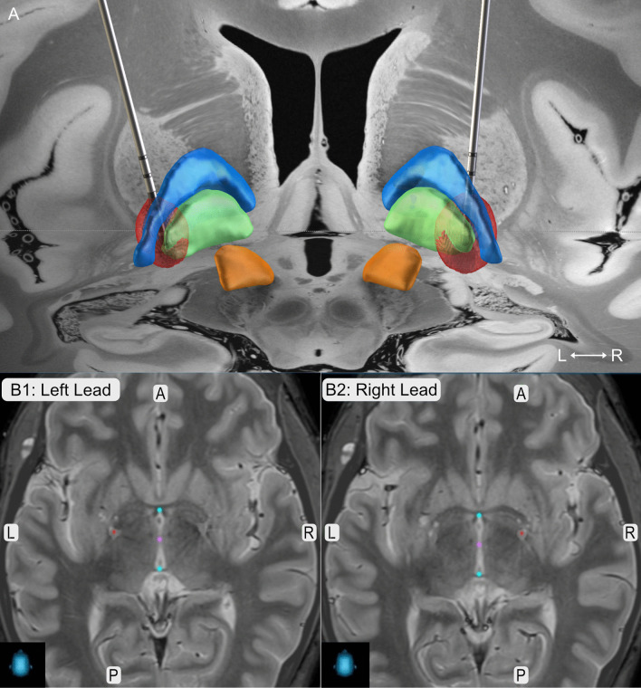
Fig. 1.
Lead Position. a Lead reconstruction in MNI ICBM 2009b space as implemented in LEAD DBS. Leads are shown in posterior view together with the globus pallidus externus (blue), globus pallidus internus (green) and the subthalamic nucleus (orange) as included in the DISTAL atlas [10, 11]. Red balls illustrate the local stimulation spread of 3-year Follow-Up stimulation parameters (Left: C+, 1-(50%), 2-(18%), 3-(16%), 4-(16%), 60 µs, 104 Hz, 4.2 mA; Right: C+, 1-(50%), 2-(18%), 3-(16%), 4-(16%), 60 µs, 104 Hz, 5.7 mA, Boston Scientific Vercise Directed lead). (B) Lead positions (red dots) as extracted fromStealthViz™, Medtronic in axial view. Coordinates in relation to AC-PC (blue dots) are x = -21.6 mm, y = -1.7 mm, z = -17.6 mm for the left lead, and x = 17.7 mm, y = 3.0 mm, z = 11.7 mm for the right lead. Abbreviations: A = anterior, L = left, P = posterior, R = right

