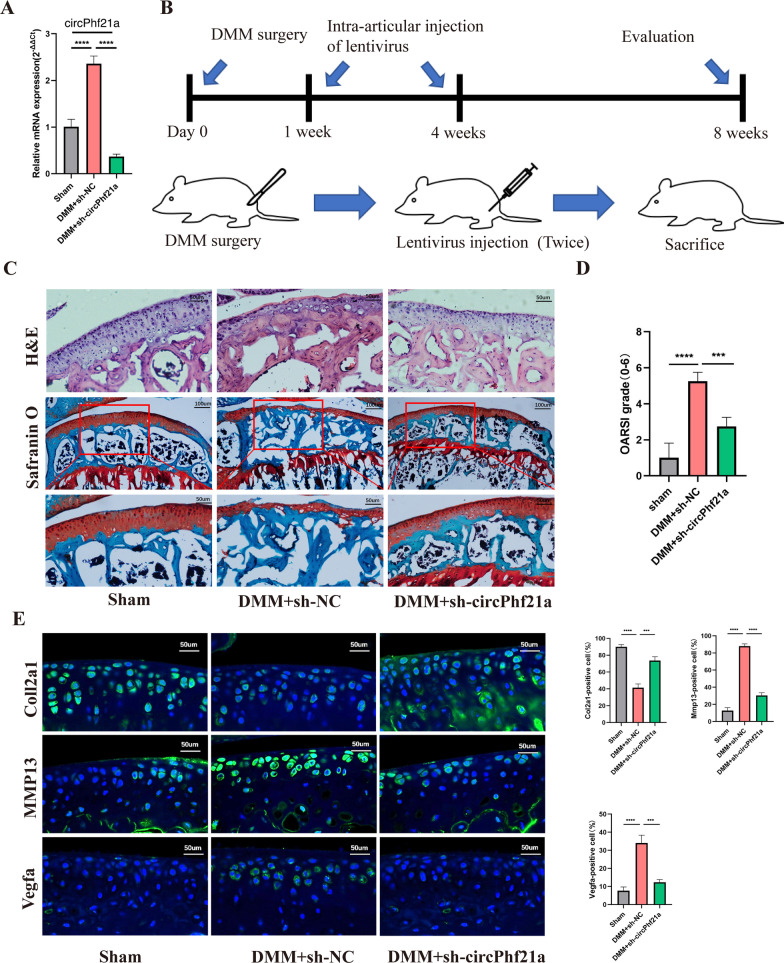
Fig. 9.
Silence of circPhf21a alleviated the progression of OA in mouse model. A RT-qPCR analysis of circPhf21a expression in knee articular cartilage from OA mice in different groups (n = 3), **p < 0.01, ****p < 0.0001. B Schematic of the time course used for the DMM-induced in vivo osteoarthritis experiments. C H&E staining was performed to observe the cell morphology and tissue integrity in the articular cartilage tissues of the mouse knee in different groups. The figures located at the top show the extent of damage and the morphology of chondrocytes in the contact area of the weight-bearing region between the medial tibial plateau and the medial femoral condyle. Likewise, the middle and bottom figures show Safranin O and fast green staining of articular cartilage tissues from mice that underwent sham or DMM surgery. Safranin O and fast green staining demonstrated OA progression through the 8-week time course in the medial tibial plateau. Scale bar = 50 or 100 μm. D OARSI scores of the medial tibial plateau of different group mice (Sham, n = 6; DMM + sh-NC, n = 6; DMM + sh-circPhf21a, n = 6). ***p < 0.001, ****p < 0.0001. E Representative images for Col2a1 (green), Mmp13 (green) and Vegfa (green) immunofluorescent staining in cartilage tissues obtained from sham or DMM mouse knees (Sham, n = 6; DMM + sh-NC, n = 6; DMM + sh-circPhf21a, n = 6). Scale bar = 50 μm. The bar graphs show quantification (%) of the Col2a1, Mmp13 or Vegfa positive cells from total cell population per field in immunofluorescent sections

