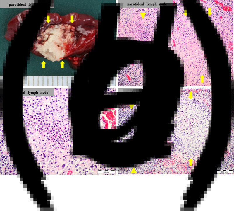
Fig. 2.
Macroscpic and microscopic lesions in the dead Eurasian beaver. (a) Focally extensive necrosis in the left parotid lymph node (arrows). (b) Left parotid lymph node; granulomatous (arrows) and necrotizing (arrowheads) lymphadenitis; H&E stain. (c) Left parotid lymph node; focal pyogranuloma; H&E stain. (d) Spleen; focal pyogranuloma (arrows) adjacent to an intact lymph follicle (arrowheads); H&E stain.

