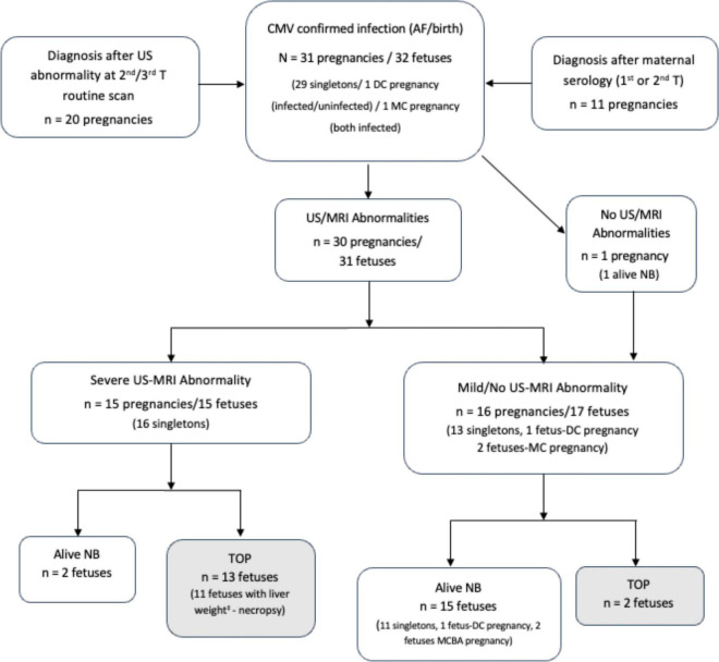
FIGURE 2.
Flowchart: Follow-up of pregnancies with confirmed fetal CMV infection. Description of outcomes according to the severity of infection, anatomopathological examination performed and liver weight at necropsy‡. DC, dichorionic; MC, monochorionic; AF, amniotic fluid; US, ultrasound; MRI, magnetic resonance imaging; TOP, termination of pregnancy; NB, newborn.

