Figure 4.
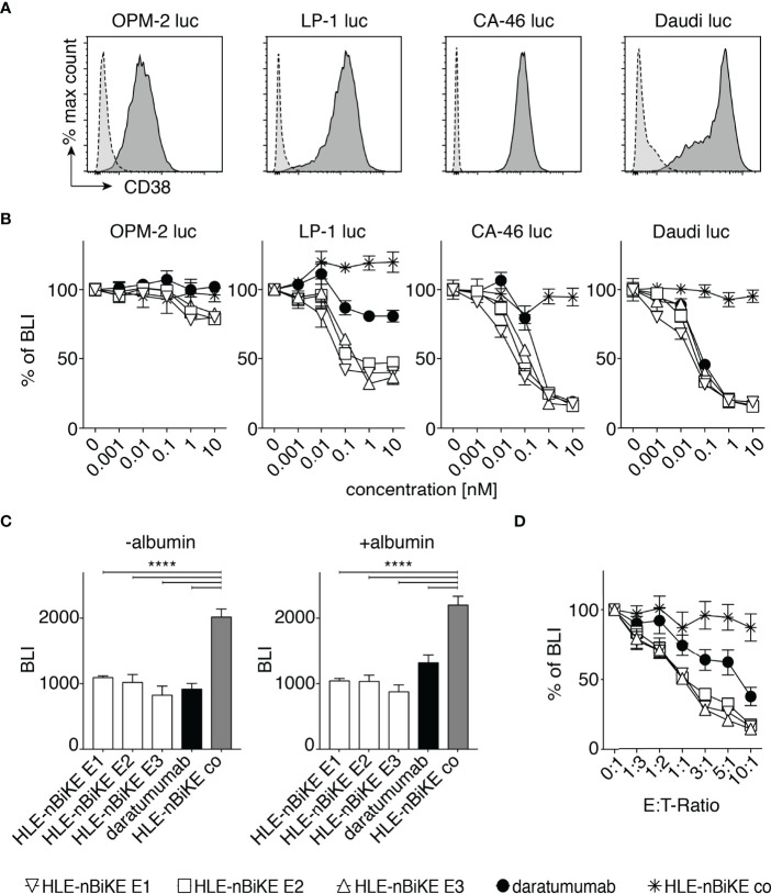
Dose and effector to target-ratio (E:T-ratio) responses of BiKE-DCC. (A) Luciferase (luc) expressing OPM-2, LP-1, CA-46, and Daudi cells were incubated with AlexaFluor 647-conjugated CD38-specific JK36 hcAb (dark grey, solid line) or isotype control s-14 hcAb (light grey, dashed line) before analysis by flow cytometry. (B) NK92 hCD16 cells were incubated an E:T-ratio of 3:1 with OPM-2 luc, LP-1 luc, CA-46, or Daudi luc cells that had been pre-incubated with the indicated concentrations of HLE-nano-BiKEs or daratumumab for 3 h at 37°C before addition of luciferin and measurement of bioluminescence-intensity (BLI) with a plate reader. Data represent mean ± SEM of triplets and are representative for three independent experiments. (C) LP-1 luc cells were incubated for 15 min with 10 nM HLE-nano-BiKEs or daratumumab in the absence or presence of 16 mg/mL albumin. Then, NK92 hCD16 cells were added at an E:T-ratio of 3:1 and incubation continued for 95 min at 37°C before addition of luciferin and measurement of BLI. Bar diagrams illustrate the mean BLI of cells after incubation with HLE-nano-BiKEs or daratumumab. One-way ANOVA was performed and Holm-Sidak-adjusted p-values are indicated (****= p < 0.0001). (D) NK92 hCD16 cells were incubated at the indicated E:T-ratios (0:1-1:10) with LP-1 luc cells that had been pre-incubated with 10 nM HLE-nano-BiKEs or daratumumab and BLI was measured after 3 h of incubation at 37°C. Values indicate the mean BLI of cells incubated with 10 nM HLE-nano-BiKEs or daratumumab as percentage of the mean BLI of cells incubated in the absence of HLE-nano-BiKEs.

