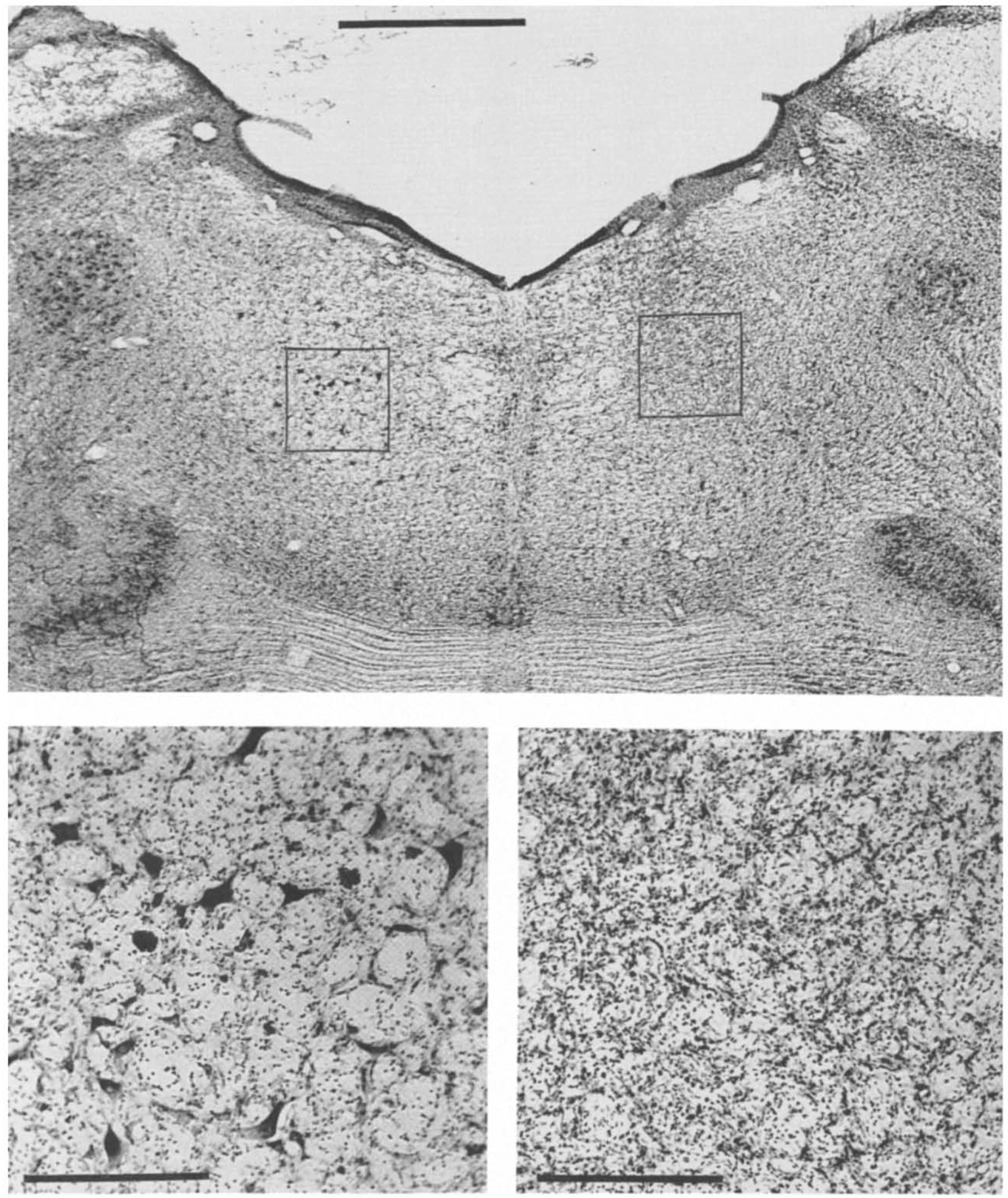
Fig. 1.

Photomicrographs of coronal sections (Cresyl violet stain) of pontine reticular formation showing IBO-lesioned (right on figure, left in brain) side and intact (left) side in cat IB-2. Bottom two sections are magnifications of lesioned and intact parts indicated on top section. Scale bar is 4 mm for top section and 0.2 mm for bottom sections.
