. 2022 Apr 27;11:e76804. doi: 10.7554/eLife.76804

Figure 4. HSV-1 capsids associate with proteins involved in type I IFN response.

Unbiased hierarchical clustered heat map showing the log2 fold changes of IFN-induced proteins (GO type-I IFN) identified from capsids-host protein sediments from cytosol of resting Mφ, or IFN-induced MφIFN macrophages. For each protein, the fold change was calculated based on their abundance (iBAQs) in V1, V0.5, and V0.1 capsids as compared to their abundance in D capsids, using a linear scale from violet being the lowest to dark green being the highest. (*) and (**) design the proteins with an FDR corrected p-value ≤ 0.05 and ≤ 0.01, respectively.

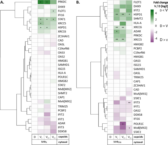
Figure 4.

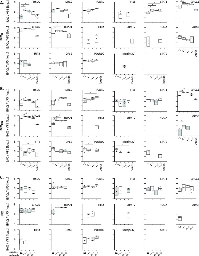
Figure 4—figure supplement 1. HSV-1 capsids binds to a few ISG proteins.

Figure 4—figure supplement 1.

Box and whisker plot of iBAQs showing the differential detection of PRKDC, DHX9, FLOT1, IFI16, STAT1, XRCC5, XRCC6, HSPD1, IFIT2, SHMT2, HLA-A, ADAR, IFIT3, OAS2, POLR1C and MxB[MX2] in D, V1, V0.5, and V0.1 capsids-host protein sediments after incubation in (A) cytosol of resting MφR macrophages, (B) IFN-induced MφIFN macrophages or (C) no cytosol. (*) design the significant binding to D or V0.1, V0.5, and V1 capsids as assessed by Welch’s t-test (two-tailed, permutation-based FDR ≤ 0.05) comparing D vs V0.1, V0.5, or V1 capsids in each cytosol separately.