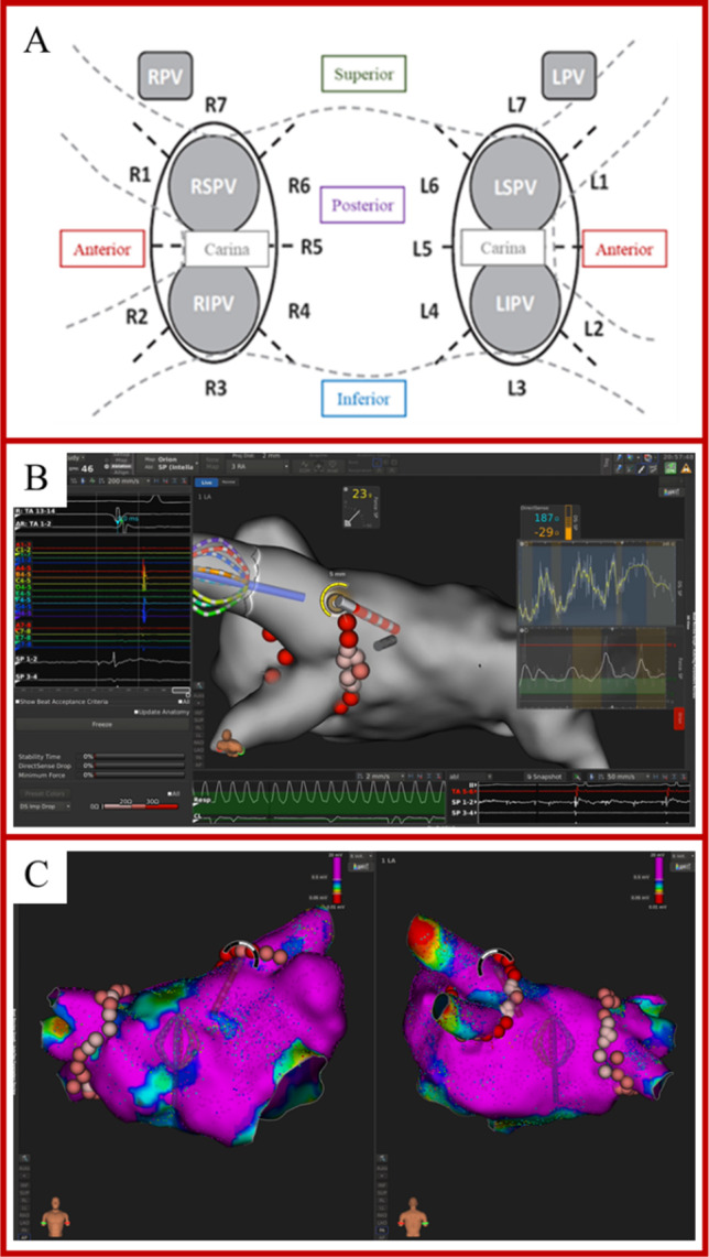
Fig. 1.

A Identification of 7 ablation sites around the right (RPV) and left (LPV) pairs of pulmonary veins. Anterior superior: R1, L1. Anterior inferior: R2, L2. Inferior: R3, L3. Posterior inferior: R4, L4. Carina: R5, L5. Posterior superior: R6, L6. Superior: R7, L7. LIPV = left inferior pulmonary vein; LSPV = left superior pulmonary vein; RIPV = right inferior pulmonary vein; RSPV = right superior pulmonary vein. B Example of visualization of CF and DirectSense™ tool on the Rhythmia™ mapping system during ablation. C Point-by-point RF delivery created contiguous ablation spots encircling the PVs. The maximal inter-lesion distance between two neighboring lesions was set ≤ 6 mm and was automatically measured through the Autotag™ software. CF settings were at the individual operator’s discretion, within the range of 5 to 40 g
