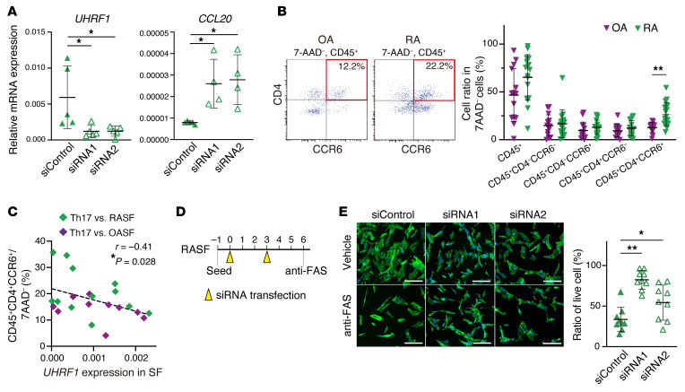
Figure 7. UHRF1 negatively regulates CCL20 expression and apoptosis resistance in RA.
(A) mRNA expression levels of UHRF1 and CCL20 in RASFs transfected with UHRF1 siRNA (n = 4–5). (B) Left, flow cytometry to measure proportion of Th17 cells (CD45+CD4+CCR6+) in OA (n = 14) and RA (n = 21) synovium tissue. Right, quantification of total CD45+ cells, CD45+CD4–CCR6– cells, CD45+CD4–CCR6+ cells, CD45+CD4+CCR6– cells, and CD45+CD4+CCR6+ cells among 7-AAD– cells. (C) Spearman’s correlation between proportion of Th17 cells and UHRF1 mRNA expression level in OASFs (n = 10) and RASFs (n = 12) obtained from synovium of the same patients. (D) Schematic protocol of consecutive UHRF1 knockdown and experimental apoptosis induction in RASFs. (E) Left, phalloidin (green) and DAPI (blue) staining of RASFs transfected twice with UHRF1 siRNA (n = 9) after treatment with 0.5 μg/mL anti-FAS antibody for 16 hours. Scale bar: 200 μm. Right, quantification of cell numbers after apoptosis induction relative to that for vehicle treatment. Mean ± SD is shown. *P < 0.05 and **P < 0.01 by ANOVA followed by Tukey’s test in A and E, and Mann-Whitney U test in B. All data were obtained from 4 to 21 independent experiments.

