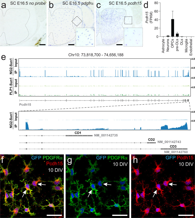
Fig. 1. Pcdh15 mRNA and protein is expressed by OPCs.
a–c Transverse spinal cord sections from embryonic day 16.5 (E16.5) mice were used to perform in situ hybridization to detect Pdgfrα or Pcdh15 mRNA. a E16.5 mouse spinal cord shows no labelling in the absence of an RNA probe. Line denotes the border of the spinal cord grey and white matter. b Pdgfrα+ OPCs are readily detected in the E16.5 spinal cord white matter. The dashed box denotes the white matter region enlarged in the lower right corner. c Image showing Pcdh15+ cells in the E16.5 mouse spinal cord. The dashed box denotes the white matter region enlarged in the lower right corner. d Quantification of Pcdh15 mRNA (in FPKM, Fragments Per Kilobase Million) in neurons and glial cells acutely purified from the early postnatal mouse brain. Graph generated using previously published and publicly available RNA sequencing data from Zhang et al.15. e Genome browser tracks of the Pcdh15 locus showing RNA-Seq alignments from nuclear RNA purified from the brains of adult Ng2-CreERT2:: Sun1-eGFP or PLP-CreERT:: Sun1-eGFP mice. Pcdh15 mRNA is highly expressed by Ng2+ OPCs and most transcripts correspond to the CD3 isoforms of Pcdh15. f–h Immunocytochemistry to detect GFP (blue), PDGFRα (green) and Pcdh15 (red) in primary OPCs generated from the early postnatal Pdgfrα-histGFP mouse cortex. Scale bars represent 60 µm (a–c) and 20 µm (f–h).

