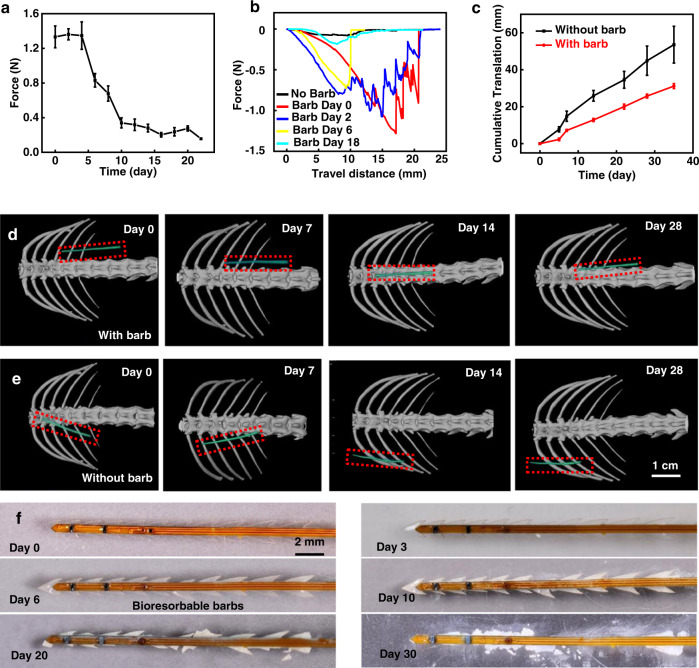
Fig. 3. Characterization of NIRS probes with bioresorbable barbs.
a Measured maximum pulling force to remove a probe inserted in a tissue phantom for different times of immersion in phosphate buffered saline (PBS) solution at 37 °C. n = 3 independent samples. All data are shown as mean ± SEM. b Representative pulling force as a function of removal distance for probes with and without barbs. c Cumulative translation distance of NIRS probes with and without barbs implanted in vivo. The measurement relies on quantitative analysis of Computed Tomography (CT) images of NIRS probes implanted subcutaneously in mice. n = 3 independent samples. All data are shown as mean ± SEM. d, e CT images showing the location of implanted NIRS probes with or without barbs respectively. All images share the same scale bar. f Images showing the process of bioresorption of barbs during soaking tests in PBS at 37 °C. Source data are provided as a Source Data file.

